MatMRI - version 2.0 released on Apr 2014
MatMRI is a library to communicate with a Philips™ MRI scanner using Matlab or Python programming language running in an external computer. The purpose of MatMRI is to use very simple instructions to initiate the communication with the MRI console and acquire images in a Matlab/Python environment, which opens quite a lot of possibilities for real-time image processing, data handling, interfacing with other devices, etc. Matlab and Python are quite deployed in many laboratories and most of users out there are knowledgeable of these platforms. MatMRI adds extra functionality such as multi-threading for an efficient data collection. MatMRI was designed to be as straightforward as possible and its use requires minimal coding.
In version 2.0, the following features were added:
And there is still new features to be added in the following months including more example codes and an even better MRI emulator to support swapping and restarting datasets.
In version 2.0, the following features were added:
- Support for Python. Thanks to native integration of .Net classes in python provided by pythonnet, PyMRI is now available. Syntax of use is practically one-to-one to Matlab code. Porting existing code should be straightforward. This Python interface is now the most used interface in our projects.
- 64-bit execution. A lot of work was done to allow MatMRI/PyMRI running in 64-bit applications. However, this should not have any impact in existing programs using matMRI. Now users should be able to use 64-bits editions of Matlab or Python.
- Support for Navigator data. Capture of navigator data is now fully tested.
- MRI Emulator. A new simulator for the generation data generated from the MR scanner was created to support emulation of magnitude, phase images and navigator data through JSON files. The emulator is capable to handle massive datasets (tens of thousands of images and navigator messages) as large as the operating system can handle. This emulator is quite useful to test code without requiring being at the MR room, which reduces considerably associated costs for the development.
And there is still new features to be added in the following months including more example codes and an even better MRI emulator to support swapping and restarting datasets.
Distribution
MatMRI is being hosted at github as a private repository and it is aimed to be freely available to researchers under coordination of Philips Healthcare. Please contact me for further enquiries
Journal article and citation
An open access journal article was accepted in the Journal of Therapeutic Ultrasound. The article showcase several applications where MatMRI is used. You can access the paper with this link. The article can be cited as: Journal of Therapeutic Ultrasound 2013, 1:7 doi:10.1186/2050-5736-1-7
Main functionality
- Asynchronous capture of individual magnitude and phase images.
- Capture of full “volumetric” data that transfers all the stacks of a sequence in a single transfer.
- Meta-data for each individual slice including echo time, position, position and orientation of slice, voxel size, etc.
- Possibility of updating the geometry information of a stack. Quite useful to change the location, orientation of a stack in a dynamic sequence.
- Possibility of pausing/resuming the scanner acquisition
Example of use
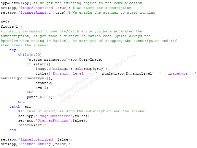
The following code is a simple example of how to collect 20 magnitude images
Application example
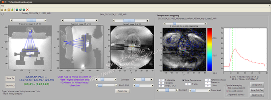
MatMRI was integrated with a small animal HIFU system and was used to capture multi-slice 2D images intended for planning as well as dynamic T1-based, single-slice images for the thermometry. In this example, a treatment with an acoustic power of 25 W was used to produce an elevation of temperature from 37◦C to 55◦C in soft tissue. For more details, please consult the journal article.
Published studies using MatMRI
- Pichardo, S,, Mougenot, C., Engler, S., Waspe, A. C.,, Drake, J. On Demand Reprogramming of MR Sequence Parameters Using MatMRI. ISMRM 24th Annual Meeting & Exhibition • 07-13 May 2016 • Singapore- Abstract 3593.
- On-Demand Dynamic Updating of the Temporal Resolution of Interleaved PRFS and T2 Temperature Mapping Methods for MR-HIFU. Engler, S., Mougenot, C., Keupp, J., Weiss, S, Heijman, E. Pichardo, S.. ISMRM 24th Annual Meeting & Exhibition • 07-13 May 2016 • Singapore- Abstract 2110
- MacDonald, M. A., Waspe, A. C., Amara, J., Pichardo, S. A Simple Scanner Control Technique for Device Localization during MRI-Guided Percutaneous Procedures. ISMRM 24th Annual Meeting & Exhibition • 07-13 May 2016 • Singapore- Abstract 3589.
- Price, K.D., Sin, V.W., Mougenot, C., Pichardo, S., Looi, T., Waspe, A.C. and Drake, J.M., 2016. Design and validation of an MR-conditional robot for transcranial focused ultrasound surgery in infants. Medical Physics, 43(9), pp.4983-4995.
- Mougenot, C., Pichardo, S., Engler, S., Waspe, A., Colas, E.C. and Drake, J.M., 2016. A rapid magnetic resonance acoustic radiation force imaging sequence for ultrasonic refocusing. Physics in Medicine and Biology, 61(15), p.5724.
- Bing, C., Nofiele, J., Staruch, R., Ladouceur-Wodzak, M., Chatzinoff, Y., Ranjan, A. and Chopra, R., 2015. Localised hyperthermia in rodent models using an MRI-compatible high-intensity focused ultrasound system.International Journal of Hyperthermia, 31(8), pp.813-822.
- Pichardo, S., Köhler, M., Lee, J. and Hynnyen, K., 2014. In vivo optimisation study for multi-baseline MR-based thermometry in the context of hyperthermia using MR-guided high intensity focused ultrasound for head and neck applications. International Journal of Hyperthermia, 30(8), pp.579-592.
- Rieck, B., Bates, D., Zhang, K., Escott, N., Mougenot, C., Pichardo, S. and Curiel, L., 2014. Focused ultrasound treatment of abscesses induced by methicillin resistant Staphylococcus aureus: Feasibility study in a mouse model. Medical physics, 41(6), p.063301.